Description
|
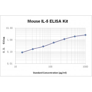
Assay Range |
15.6–1000 pg/mL |
|
Sensitivity |
2.0 pg/mL |
|
Specificity |
No cross-reaction with other related substances detected |
|
Size |
96T |
|
Storage |
Store at 2 – 8ºC. Keep reconstituted standard and detection Ab at -20 ºC |
|
Assay Principle |
Sandwich ELISA |
|
Sample Volume |
100 µL final volume, dilution factor varies on samples |
|
Sample Type |
serum, plasma or cell culture supernatant |
|
Detection Method |
Chromogenic |
Kit Components
1. Recombinant Mouse IL-5 standard: 2 vials
2. One 96-well plate coated with Mouse IL-5 Ab
3. Sample diluent buffer: 12 mL – 1
4. Detection antibody: 130 µL, dilution 1:100
5. Streptavidin-HRP: 130 µL, dilution 1:100
6. Antibody diluent buffer: 12 mL x1
7. Streptavidin-HRP diluent buffer: 12 mL x1
8. TMB developing agent: 10 mL x1
9. Stop solution: 10 mL x1
10. Washing solution (20x): 25 mL x1
Background
Interleukin 5 (IL-5), also known as B-cell differentiation factor I, Eosinophil differentiation factor, T-cell replacing factor (TRF), is a secreted glycoprotein that belongs to the α-helical group of cytokines including IL-3, IL-5, and GM-CSF. IL-5 is primarily expressed by CD4+ Th2 cells, while activated eosinophils and mast cells can also secreted it with lower amounts. Native mouse IL-5 is a disulfide-linked homodimeric glycoprotein. Mouse IL-5 is synthesized as a 133 amino acid (aa) precursor protein composed of a 20aa hydrophobic signal peptide and a 113 aa mature protein. Mouse IL-5 is 70% identical to human IL-5 and exhibits equal activity with human IL-5 on human cell lines. Mouse IL-5 binds to a heterodimeric receptor complex consisting of an α and a β subunit to trigger a series of biological activities in target cells. The α subunit binds IL-5 specifically with intermediate affinity. The β subunit, also known as AIC2B, is a common subunit (βc) shared by IL-3, IL-5 or GM-CSF. Apart from the membrane-bound form of mouse IL-5Rα, soluble isoforms of IL-5 Rα have also been identified and shown to be an IL-5 antagonist. Mouse IL-5 Rα expression is restricted to eosinophils, B cells and mast cells. The expression of the βc subunit, however, is detected on various lineages of hematopoietic cells.