Description
Overview
Long Name |
Antibody Type |
Antibody Isotype |
Host |
Species Reactivity |
Validated Applications |
Purification |
| perforin 1 (pore forming protein) | Polyclonal | IgG | Rabbit | Human | WB | Immunogen affinity purified. |
Immunogen |
||||||
| E.coli-derived human Perforin recombinant protein (Position: E175-W555). Human Perforin shares 68% amino acid (aa) sequence identity with both mouse and rat Perforin. | ||||||
Properties
Form |
Lyophilized |
Size |
100 µg/vial |
Contents |
Antibody is lyophilized with 5 mg BSA, 0.9 mg NaCl, 0.2 mg Na2HPO4, 0.05 mg NaN3. *carrier free antibody available upon request. |
Concentration |
Reconstitute with 0.2 mL sterile dH2O (500 µg/ml final concentration). |
Storage |
At -20 °C for 12 months, as supplied. Store reconstituted antibody at 2-8 °C for one month. For long-term storage, aliquot and store at -20 °C. Avoid repeated freezing and thawing. |
Additional Information Regarding the Antigen
Gene |
PRF1 |
Protein |
Perforin-1 |
Uniprot ID |
P14222 |
Function |
Plays a key role in secretory granule-dependent cell death, and in defense against virus-infected or neoplastic cells. Plays an important role in killing other cells that are recognized as non-self by the immune system, e.g. in transplant rejection or some forms of autoimmune disease. Can insert into the membrane of target cells in its calcium-bound form, oligomerize and form large pores. Promotes cytolysis and apoptosis of target cells by facilitating the uptake of cytotoxic granzymes. |
Tissue Specificity |
|
Sub-cellular localization |
Cytoplasmic granule lumen. Secreted. Cell membrane; Multi-pass membrane protein. Endosome lumen. Note: Stored in cytoplasmic granules of cytolytic T-lymphocytes and secreted into the cleft between T-lymphocyte and target cell. Inserts into the cell membrane of target cells and forms pores. Membrane insertion and pore formation requires a major conformation change. May be taken up via endocytosis involving clathrin-coated vesicles and accumulate in a first time in large early endosomes. |
Sequence Similarities |
Belongs to the complement C6/C7/C8/C9 family. |
Aliases |
Cytolysin antibody|FLH2 antibody|HPLH2 antibody|Lymphocyte pore forming protein antibody|Lymphocyte pore-forming protein antibody|MGC65093 antibody|P1 antibody|PERF_HUMAN antibody|perforin 1 (pore forming protein) antibody|Perforin 1 antibody|Perforin 1 precursor antibody|Perforin 1 preforming protein antibody|Perforin-1 antibody|PFP antibody|PGFL antibody|PIGF antibody|PIGF-2 antibody|PLGF antibody|Pore forming protein antibody|PRF 1 antibody|PRF1 antibody|SHGC-10760 antibody |
Application Details
| Application | Concentration* | Species | Validated Using** |
| Western blot | 0.1-0.5μg/ml | Human | AssaySolutio’s ECL kit |
AssaySolution recommends Rabbit Chemiluminescent WB Detection Kit (AKIT001B) for Western blot. *Blocking peptide can be purchased at $65. Contact us for more information

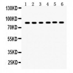
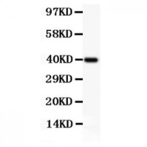
Anti-Perforin antibody, ASA-B1499–1.jpg
All lanes: Anti Perforin (ASA-B1499) at 0.5ug/ml
WB: Recombinant Human Perforin Protein 0.5ng
Predicted bind size: 40KD
Observed bind size: 40KD
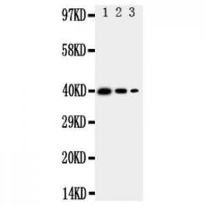
All lanes: Anti Perforin (ASA-B1499) at 0.5ug/ml
WB: Recombinant Human Perforin Protein 0.5ng
Predicted bind size: 40KD
Observed bind size: 40KD

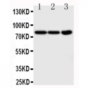
Anti-Perforin antibody, ASA-B1499–2.jpg
All lanes: Anti Perforin (ASA-B1499) at 0.5ug/ml
Lane 1: HELA Whole Cell Lysate at 40ug
Lane 2: COLO320 Whole Cell Lysate at 40ug
Lane 3: HEPG2 Whole Cell Lysate at 40ug
Predicted bind size: 61KD
Observed bind size: 61KD
All lanes: Anti Perforin (ASA-B1499) at 0.5ug/ml
Lane 1: HELA Whole Cell Lysate at 40ug
Lane 2: COLO320 Whole Cell Lysate at 40ug
Lane 3: HEPG2 Whole Cell Lysate at 40ug
Predicted bind size: 61KD
Observed bind size: 61KD