Description
Overview
Long Name |
Antibody Type |
Antibody Isotype |
Host |
Species Reactivity |
Validated Applications |
Purification |
| ATP synthase, H+ transporting, mitochondrial Fo complex, subunit d | Polyclonal | IgG | Rabbit | Human, Mouse, Rat | IHC-P, ICC, WB | Immunogen affinity purified. |
Immunogen |
||||||
| E.coli-derived human ATP5H recombinant protein (Position: A2-L161). Human ATP5H shares 81% and 78% amino acid (aa) sequence identity with mouse and rat ATP5H, respectively. | ||||||
Properties
Form |
Lyophilized |
Size |
100 µg/vial |
Contents |
Antibody is lyophilized with 5 mg BSA, 0.9 mg NaCl, 0.2 mg Na2HPO4, 0.05 mg NaN3. *carrier free antibody available upon request. |
Concentration |
Reconstitute with 0.2 mL sterile dH2O (500 µg/ml final concentration). |
Storage |
At -20 °C for 12 months, as supplied. Store reconstituted antibody at 2-8 °C for one month. For long-term storage, aliquot and store at -20 °C. Avoid repeated freezing and thawing. |
Additional Information Regarding the Antigen
Gene |
ATP5H |
Protein |
ATP synthase subunit d, mitochondrial |
Uniprot ID |
O75947 |
Function |
Mitochondrial membrane ATP synthase (F(1)F(0) ATP synthase or Complex V) produces ATP from ADP in the presence of a proton gradient across the membrane which is generated by electron transport complexes of the respiratory chain. F-type ATPases consist of two structural domains, F(1) – containing the extramembraneous catalytic core, and F(0) – containing the membrane proton channel, linked together by a central stalk and a peripheral stalk. During catalysis, ATP synthesis in the catalytic domain of F(1) is coupled via a rotary mechanism of the central stalk subunits to proton translocation. Part of the complex F(0) domain and the peripheric stalk, which acts as a stator to hold the catalytic alpha(3)beta(3) subcomplex and subunit a/ATP6 static relative to the rotary elements. |
Tissue Specificity |
|
Sub-cellular localization |
Mitochondrion. Mitochondrion inner membrane. |
Sequence Similarities |
|
Aliases |
ATP synthase D chain mitochondrial antibody|ATP synthase H+ transporting mitochondrial F1F0 subunit antibody|ATP synthase H+ transporting mitochondrial F1F0 subunit d antibody|ATP synthase subunit d antibody|ATP synthase subunit d, mitochondrial antibody|ATP synthase, H+ transporting, mitochondrial F0 complex, subunit d antibody|ATP5H antibody|ATP5H_HUMAN antibody|ATP5JD antibody|ATPase subunit d antibody|ATPQ antibody|mitochondrial antibody|My032 protein antibody |
Application Details
| Application | Concentration* | Species | Validated Using** |
| Western blot | 0.1-0.5μg/ml | Human, Mouse, Rat | AssaySolutio’s ECL kit |
| Immunohistochemistry(Paraffin-embedded Section) | 0.5-1μg/ml | Human, Mouse, Rat | AssaySolutio’s IHC/ICC Detection kit |
| Immunocytochemistry | 0.5-1μg/ml | Human | AssaySolutio’s IHC/ICC Detection kit |
AssaySolution recommends Rabbit Chemiluminescent WB Detection Kit (AKIT001B) for Western blot, and Rabbit Peroxidase IHC/ICC Detection Kit (AKIT002B) for IHC(P) and ICC. *Blocking peptide can be purchased at $65. Contact us for more information

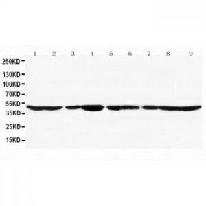
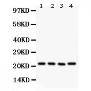
Anti- ATP5H antibody, ASA-B0174, Western blotting
All lanes: Anti ATP5H (ASA-B0174) at 0.5ug/ml
Lane 1: Rat Brain Tissue Lysate at 50ug
Lane 2: Mouse Brain Tissue Lysate at 50ug
Lane 3: Human Placenta Tissue Lysate at 50ug
Lane 4: HELA Whole Cell Lysate at 40ug
Predicted band size: 22KD
Observed band size: 22KD
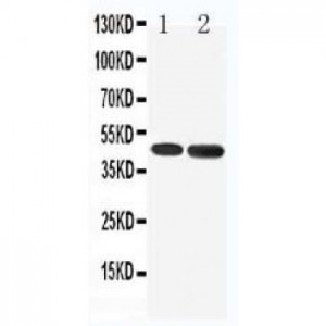
All lanes: Anti ATP5H (ASA-B0174) at 0.5ug/ml
Lane 1: Rat Brain Tissue Lysate at 50ug
Lane 2: Mouse Brain Tissue Lysate at 50ug
Lane 3: Human Placenta Tissue Lysate at 50ug
Lane 4: HELA Whole Cell Lysate at 40ug
Predicted band size: 22KD
Observed band size: 22KD

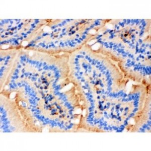
Anti- ATP5H antibody, ASA-B0174,IHC(P)
Mouse Intestine Tissue
Mouse Intestine Tissue

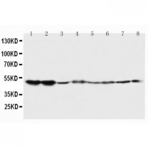
Anti- ATP5H antibody, ASA-B0174,IHC(P)
Rat Intestine Tissue
Rat Intestine Tissue

Anti- ATP5H antibody, ASA-B0174,IHC(P)
Human Mammary Cancer Tissue