Description
Overview
Long Name |
Antibody Type |
Antibody Isotype |
Host |
Species Reactivity |
Validated Applications |
Purification |
| dystrophin | Polyclonal | IgG | Rabbit | Human, Mouse, Rat | IHC-P, WB | Immunogen affinity purified. |
Immunogen |
||||||
| E.coli-derived human Dystrophin recombinant protein (Position: H3076-D3404). Human Dystrophin shares 100% amino acid (aa) sequence identity with mouse Dystrophin. | ||||||
Properties
Form |
Lyophilized |
Size |
100 µg/vial |
Contents |
Antibody is lyophilized with 5 mg BSA, 0.9 mg NaCl, 0.2 mg Na2HPO4, 0.05 mg NaN3. *carrier free antibody available upon request. |
Concentration |
Reconstitute with 0.2 mL sterile dH2O (500 µg/ml final concentration). |
Storage |
At -20 °C for 12 months, as supplied. Store reconstituted antibody at 2-8 °C for one month. For long-term storage, aliquot and store at -20 °C. Avoid repeated freezing and thawing. |
Additional Information Regarding the Antigen
Gene |
DMD |
Protein |
Dystrophin |
Uniprot ID |
P11532 |
Function |
Anchors the extracellular matrix to the cytoskeleton via F-actin. Ligand for dystroglycan. Component of the dystrophin- associated glycoprotein complex which accumulates at the neuromuscular junction (NMJ) and at a variety of synapses in the peripheral and central nervous systems and has a structural function in stabilizing the sarcolemma. Also implicated in signaling events and synaptic transmission. |
Tissue Specificity |
Expressed in muscle fibers accumulating in the costameres of myoplasm at the sarcolemma. Expressed in brain, muscle, kidney, lung and testis. Isoform 5 is expressed in heart, brain, liver, testis and hepatoma cells. Most tissues contain transcripts of multiple isoforms, however only isoform 5 is detected in heart and liver. |
Sub-cellular localization |
Cell membrane, sarcolemma; Peripheral membrane protein; Cytoplasmic side. Cytoplasm, cytoskeleton. Cell junction, synapse, postsynaptic cell membrane . Note: In muscle cells, sarcolemma localization requires the presence of ANK2, while localization to costameres requires the presence of ANK3. Localizes to neuromuscular junctions (NMJs) in the presence of ANK2 (By similarity). |
Sequence Similarities |
Contains 2 CH (calponin-homology) domains. |
Aliases |
Apo dystrophin antibody|BMD antibody|CMD3B antibody|DMD antibody|DMD_HUMAN antibody|Duchenne muscular dystrophy protein antibody|DXS142 antibody|DXS164 antibody|DXS206 antibody|DXS230 antibody|DXS239 antibody|DXS268 antibody|DXS269 antibody|DXS270 antibody|DXS272 antibody|Dystrophin antibody|Muscular dystrophy Duchenne and Becker types antibody |
Application Details
| Application | Concentration* | Species | Validated Using** |
| Western blot | 0.1-0.5μg/ml | Human, Mouse, Rat | AssaySolutio’s ECL kit |
| Immunohistochemistry(Paraffin-embedded Section) | 0.5-1μg/ml | Human, Mouse, Rat | AssaySolutio’s IHC/ICC Detection kit |
AssaySolution recommends Rabbit Chemiluminescent WB Detection Kit (AKIT001B) for Western blot, and Rabbit Peroxidase IHC/ICC Detection Kit (AKIT002B) for IHC(P). *Blocking peptide can be purchased at $65. Contact us for more information

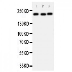
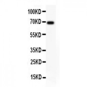
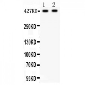
Anti- Dystrophin antibody, ASA-B0616, Western blotting
All lanes: Anti Dystrophin (ASA-B0616) at 0.5ug/ml
Lane 1: SMMC Whole Cell Lysate at 40ug
Lane 2: HEPA Whole Cell Lysate at 40ug
Predicted bind size: 427KD
Observed bind size: 427KD
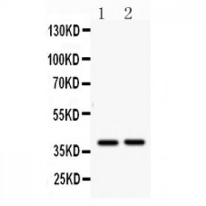
All lanes: Anti Dystrophin (ASA-B0616) at 0.5ug/ml
Lane 1: SMMC Whole Cell Lysate at 40ug
Lane 2: HEPA Whole Cell Lysate at 40ug
Predicted bind size: 427KD
Observed bind size: 427KD

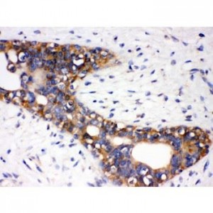
Anti- Dystrophin antibody, ASA-B0616, IHC(P)
IHC(P): Mouse Brain Tissue
IHC(P): Mouse Brain Tissue

Anti- Dystrophin antibody, ASA-B0616, IHC(P)
IHC(P): Rat Cardiac Muscle Tissue
IHC(P): Rat Cardiac Muscle Tissue