Description
Overview
Long Name |
Antibody Type |
Antibody Isotype |
Host |
Species Reactivity |
Validated Applications |
Purification |
| cadherin 1, type 1, E-cadherin (epithelial) | Polyclonal | IgG | Rabbit | Human | ELISA, IHC-P, IHC-F, WB | Immunogen affinity purified. |
Immunogen |
||||||
| E.coli-derived human E Cadherin recombinant protein (Position: A286-A703). Human E Cadherin shares 79.7% and 80.9% amino acid (aa) sequence identity with mouse and rat E Cadherin, respectively. | ||||||
Properties
Form |
Lyophilized |
Size |
100 µg/vial |
Contents |
Antibody is lyophilized with 5 mg BSA, 0.9 mg NaCl, 0.2 mg Na2HPO4, 0.05 mg NaN3. *carrier free antibody available upon request. |
Concentration |
Reconstitute with 0.2 mL sterile dH2O (500 µg/ml final concentration). |
Storage |
At -20 °C for 12 months, as supplied. Store reconstituted antibody at 2-8 °C for one month. For long-term storage, aliquot and store at -20 °C. Avoid repeated freezing and thawing. |
Additional Information Regarding the Antigen
Gene |
CDH1 |
Protein |
Cadherin-1 |
Uniprot ID |
P12830 |
Function |
Cadherins are calcium-dependent cell adhesion proteins. They preferentially interact with themselves in a homophilic manner in connecting cells; cadherins may thus contribute to the sorting of heterogeneous cell types. CDH1 is involved in mechanisms regulating cell-cell adhesions, mobility and proliferation of epithelial cells. Has a potent invasive suppressor role. It is a ligand for integrin alpha-E/beta-7. |
Tissue Specificity |
Non-neural epithelial tissues. |
Sub-cellular localization |
Cell junction. Cell membrane; Single-pass type I membrane protein. Endosome. Golgi apparatus, trans-Golgi network. Note: Colocalizes with DLGAP5 at sites of cell-cell contact in intestinal epithelial cells. Anchored to actin microfilaments through association with alpha-, beta- and gamma- catenin. Sequential proteolysis induced by apoptosis or calcium influx, results in translocation from sites of cell-cell contact to the cytoplasm. Colocalizes with RAB11A endosomes during its transport from the Golgi apparatus to the plasma membrane. |
Sequence Similarities |
Contains 5 cadherin domains. |
Aliases |
Arc 1 antibody|CADH1_HUMAN antibody|Cadherin 1 antibody|cadherin 1 type 1 E-cadherin antibody|Cadherin1 antibody|CAM 120/80 antibody|CD 234 antibody|CD 324 antibody|CD324 antibody|CD324 antigen antibody|CDH1 antibody|CDHE antibody|E-Cad/CTF3 antibody|E-cadherin antibody|ECAD antibody|Epithelial cadherin antibody|epithelial calcium dependant adhesion protein antibody|LCAM antibody|Liver cell adhesion molecule antibody|UVO antibody| Uvomorulin antibody |
Application Details
| Application | Concentration* | Species | Validated Using** |
| Western blot | 0.1-0.5μg/ml | Human | AssaySolutio’s ECL kit |
| Immunohistochemistry(Paraffin-embedded Section) | 0.5-1μg/ml | Human | AssaySolutio’s IHC/ICC Detection kit |
| Immunohistochemistry(Frozen Section) | 0.5-1μg/ml | Human | AssaySolutio’s IHC/ICC Detection kit |
| ELISA | 0.1-0.5μg/ml | Human | Sandwich ELISA format |
AssaySolution recommends Rabbit Chemiluminescent WB Detection Kit (AKIT001B) for Western blot, and Rabbit Peroxidase IHC/ICC Detection Kit (AKIT002B) for IHC(P) and ICC. *Blocking peptide can be purchased at $65. Contact us for more information

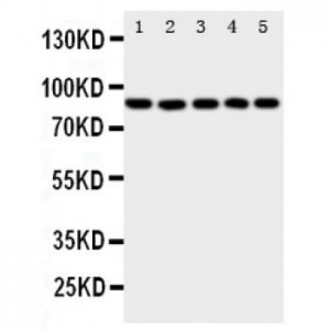
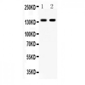
Anti- E Cadherin antibody, ASA-B0618, Western blotting
All lanes: Anti E Cadherin (ASA-B0618) at 0.5ug/ml
Lane 1: Human Placenta Tissue Lysate at 50ug
Lane 2: HELA Whole Cell Lysate at 40ug
Predicted bind size: 140KD
Observed bind size: 140KD
All lanes: Anti E Cadherin (ASA-B0618) at 0.5ug/ml
Lane 1: Human Placenta Tissue Lysate at 50ug
Lane 2: HELA Whole Cell Lysate at 40ug
Predicted bind size: 140KD
Observed bind size: 140KD

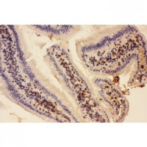
Anti- E Cadherin antibody, ASA-B0618, IHC(P)
IHC(P): Human Placenta Tissue
IHC(P): Human Placenta Tissue

Anti- E Cadherin antibody, ASA-B0618, IHC(F)
IHC(F): Human Placenta Tissue
IHC(F): Human Placenta Tissue