Description
Overview
Long Name |
Antibody Type |
Antibody Isotype |
Host |
Species Reactivity |
Validated Applications |
Purification |
| clock circadian regulator | Polyclonal | IgG | Rabbit | Human, Mouse, Rat | IHC-P, WB | Immunogen affinity purified. |
Immunogen |
||||||
| A synthetic peptide corresponding to a sequence at the N-terminus of human KAT13D/CLOCK (75-109aa QKSIDFLRKHKEITAQSDASEIRQDWKPTFLSNEE) , different from the related mouse sequence by one amino acid, and identical to the related rat sequence. | ||||||
Properties
Form |
Lyophilized |
Size |
100 µg/vial |
Contents |
Antibody is lyophilized with 5 mg BSA, 0.9 mg NaCl, 0.2 mg Na2HPO4, 0.05 mg NaN3. *carrier free antibody available upon request. |
Concentration |
Reconstitute with 0.2 mL sterile dH2O (500 µg/ml final concentration). |
Storage |
At -20 °C for 12 months, as supplied. Store reconstituted antibody at 2-8 °C for one month. For long-term storage, aliquot and store at -20 °C. Avoid repeated freezing and thawing. |
Additional Information Regarding the Antigen
Gene |
CLOCK |
Protein |
Circadian locomoter output cycles protein kaput |
Uniprot ID |
O15516 |
Function |
Transcriptional activator which forms a core component of the circadian clock. The circadian clock, an internal time- keeping system, regulates various physiological processes through the generation of approximately 24 hour circadian rhythms in gene expression, which are translated into rhythms in metabolism and behavior. It is derived from the Latin roots ‘circa’ (about) and ‘diem’ (day) and acts as an important regulator of a wide array of physiological functions including metabolism, sleep, body temperature, blood pressure, endocrine, immune, cardiovascular, and renal function. Consists of two major components: the central clock, residing in the suprachiasmatic nucleus (SCN) of the brain, and the peripheral clocks that are present in nearly every tissue and organ system. Both the central and peripheral clocks can be reset by environmental cues, also known as Zeitgebers (German for ‘timegivers’). The predominant Zeitgeber for the central clock is light, which is sensed by retina and signals directly to the SCN. The central clock entrains the peripheral clocks through neuronal and hormonal signals, body temperature and feeding-related cues, aligning all clocks with the external light/dark cycle. Circadian rhythms allow an organism to achieve temporal homeostasis with its environment at the molecular level by regulating gene expression to create a peak of protein expression once every 24 hours to control when a particular physiological process is most active with respect to the solar day. Transcription and translation of core clock components (CLOCK, NPAS2, ARNTL/BMAL1, ARNTL2/BMAL2, PER1, PER2, PER3, CRY1 and CRY2) plays a critical role in rhythm generation, whereas delays imposed by post-translational modifications (PTMs) are important for determining the period (tau) of the rhythms (tau refers to the period of a rhythm and is the length, in time, of one complete cycle). A diurnal rhythm is synchronized with the day/night cycle, while the ultradian and infradian rhythms have a period shorter and longer than 24 hours, respectively. Disruptions in the circadian rhythms contribute to the pathology of cardiovascular diseases, cancer, metabolic syndromes and aging. A transcription/translation feedback loop (TTFL) forms the core of the molecular circadian clock mechanism. Transcription factors, CLOCK or NPAS2 and ARNTL/BMAL1 or ARNTL2/BMAL2, form the positive limb of the feedback loop, act in the form of a heterodimer and activate the transcription of core clock genes and clock-controlled genes (involved in key metabolic processes), harboring E-box elements (5′-CACGTG-3′) within their promoters. The core clock genes: PER1/2/3 and CRY1/2 which are transcriptional repressors form the negative limb of the feedback loop and interact with the CLOCK |
Tissue Specificity |
Hair follicles (at protein level). Expressed in all tissues examined including spleen, thymus, prostate, testis, ovary, small intestine, colon, leukocytes, heart, brain, placenta, lung, liver, skeletal muscle, kidney and pancreas. Highest levels in testis and skeletal muscle. Low levels in thymus, lung and liver. Expressed in all brain regions with highest levels in cerebellum. Highly expressed in the suprachiasmatic nucleus (SCN). |
Sub-cellular localization |
Nucleus. |
Sequence Similarities |
Contains 1 bHLH (basic helix-loop-helix) domain. |
Aliases |
bHLHe8 antibody|Circadian locomoter output cycles kaput protein antibody|Circadian locomoter output cycles protein kaput antibody|Circadian Locomotor Output Cycles Kaput antibody|Circadium Locomotor Output Cycles Kaput antibody|Class E basic helix-loop-helix protein 8 antibody|CLOCK antibody| Clock circadian regulator antibody|Clock homolog antibody|Clock protein antibody|CLOCK_HUMAN antibody|hCLOCK antibody|KIAA0334 antibody |
Application Details
| Application | Concentration* | Species | Validated Using** |
| Western blot | 0.1-0.5μg/ml | Human, Mouse, Rat | AssaySolutio’s ECL kit |
| Immunohistochemistry(Paraffin-embedded Section) | 0.5-1μg/ml | Human, Mouse, Rat | AssaySolutio’s IHC/ICC Detection kit |
AssaySolution recommends Rabbit Chemiluminescent WB Detection Kit (AKIT001B) for Western blot, and Rabbit Peroxidase IHC/ICC Detection Kit (AKIT002B) for IHC(P). *Blocking peptide can be purchased at $65. Contact us for more information

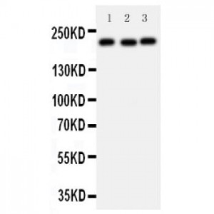
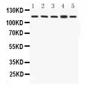
Anti- KAT13D/CLOCK antibody, ASA-B1102, Western blotting
All lanes: Anti KAT13D/CLOCK (ASA-B1102) at 0.5ug/ml
Lane 1: Rat Skeletal Muscle Tissue Lysate at 50ug
Lane 2: Mouse Skeletal Muscle Tissue Lysate at 50ug
Lane 3: NIH3T3 Whole Cell Lysate at 40ug
Lane 4: A549 Whole Cell Lysate at 40ug
Lane 5: MCF7 Whole Cell Lysate at 40ug
Predicted bind size: 95KD
Observed bind size: 115KD
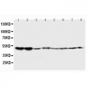

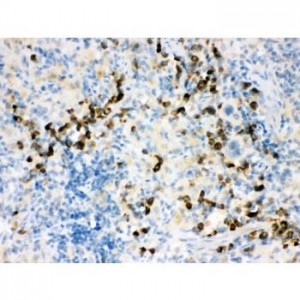
Anti- KAT13D/CLOCK antibody, ASA-B1102, IHC(P)
IHC(P): Mouse Skeletal Muscle Tissue
IHC(P): Mouse Skeletal Muscle Tissue

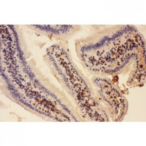
Anti- KAT13D/CLOCK antibody, ASA-B1102, IHC(P)
IHC(P): Rat Testis Tissue
IHC(P): Rat Testis Tissue